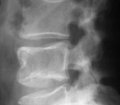
Somehow, radiographic examination is still a controversial subject in the practice of chiropractic. Many chiropractic techniques and management strategies rely on assessment protocols involving palpation, leg-checks, manual muscle testing, thermography, SEMG and others to detect and locate vertebral subluxation. While various assessment protocols may aid chiropractors in delivering beneficial care, a radiographic examination is vital for a varierty of reasons.
1. Determining the safety and appropriateness of chiropractic care.
2. The detection and characterization of vertebral subluxations
3. Determination of congenital and developmental anomalies which may affect the selection of chiropractic techniques
4. Ruling out conditions which may contraindicate certain chiropractic adjusting methods.
5. Radiography may disclose conditions requiring referral to another type of health care provider.
CLICK HERE for Dr. Kent's paper
Souza, which is a required textbook in most chiropractic programs in the US, suggests that patients who present with low back pain should be ruled out for serious conditions such as canal stenosis, spondylolisthesis, ankylosing spondylitis, Reiter’s syndrome, multiple myeloma, metastatic carcinoma, and abdominal aortic aneurysm (AAA).
As an example, AAA causes death in 80% of subjects when left untreated. In 2009, AAAs resulted in approximately 15,000 deaths. Chiropractic offices may encounter AAAs more often than expected since it often presents as LBP until rupture. However, AAA can be asymptomatic before rupture and a ruptured AAA can end in death. For this reason, every chiropractor should prepare for the possibility of AAAs or any number of other serious conditions presenting as back pain or neck pain for that matter.
This emphasizes the need for x-ray examination and analysis.
Patients should be well-informed about the risks and benefits of radiation. However, this requires an up-to- date understanding of that risk-benefit analysis using the available evidence. For many years, the linear no-threshold (LNT) dose-response model dominated healthcare regarding exposure to radiation. The LNT states that exposure to radiation is related to harmful carcinogenic effects and can be “linearly extrapolated to much lower doses that are encountered in diagnostic medical procedures”.
The alarming conclusions from this theory has echoed through global media, provoking widespread public concern. However, the LNT has been invalidated by new recent studies suggesting “that low dose radiation would decrease cancer risk thanks to the enhancement of immune system response”.
Zanzonico et. al quantitatively compare actual mortality and morbidity data and determined that foregoing radiographic imaging have led to misdiagnosis, missed diagnosis, suboptimal care, and poor outcomes which exceed the hypothetical radiogenic risk of a radiographic examination.
Good healthcare practice includes a proper informed consent about the benefits and risks of health procedures. More and more scientific evidence is ruling out the LNT and supporting the radiation hormesis (or radiation homeostasis) theory. Radiation hormesis states that low doses of radiation stimulate immune responses and repair mechanisms that protect against disease, which would otherwise be inactivated.
Luckey states, "for every thousand cancer mortalities predicted by the linear models (LNT), there will be a thousand decreased cancer mortalities and ten thousand persons with improved quality of life”. As noted by Siegel, it’s time to accept the evidence and call for an ending to radiophobia.
In a commentary, reviewing the literature on the phantom risks of radiographic imaging, Oakley et. al state:
There is essentially no scientifically demonstrable risk to the given patient. Further, follow-up radiographs to monitor response to treatment as required by some technique protocols, also provide negligible risk. Chiropractic radiologists and those pressing for more restrictive guidelines to dictate restraint in clinical x-ray use should know these efforts are not supported by scientific evidence and are anti-progressive and costly. Efforts to limit the use of x-ray in chiropractic clinical practice by influencing the education of the student at chiropractic college or by devising more restrictive practice guidelines to restrict the chiropractor in practice should be ceased. It is now time for radiologists to reconsider or even reverse their opinions on the risks of x-ray usage in practice.
Siegel states: "The low-dose radiation of [radiographic] imaging has no documented pathway to harm,” whereas an unsubstantiated refrain from the critical valid, reliable, and objective findings from radiographic examinations and analyses most assuredly does.
REFERENCES:
1. Souza TA. Differential diagnosis and management for the chiropractor protocols and algorithms. 4 th ed. Massachusetts: Jones and Bartlett; 2009.
2. Bloomer LDS, Bown MJ. Sexual dimorphism of abdominal aortic aneurysms: A striking example of “male disadvantage” in cardiovascular disease. Eur J Vasc Endovasc Surg. 2012 Jun;22:1-7.
3. Laurent A, Morel E, editors. Aneurysms: Types, risks, formation and treatment. New York: Nova Science Publishers; 2009.
4. Kauppila LI, McAlindon T, Evans S, Wilson PWF, Kiel D, FelsonDT. Disc degeneration/back pain and calicification of the abdominal aorta. Spine. 1997;22:1642-9.
5. Zanzonico PB. The Neglected Side of the Coin: Quantitative Benefit-risk Analyses in Medical Imaging. Health Phys. 2016 Mar;110(3):301-4.
6. Tomà P, Cannatà V, Genovese E, Magistrelli A, Granata C. Radiation exposure in diagnostic imaging: wisdom and prudence, but still a lot to understand. Radiol Med. 2017 Mar;122(3):215-220.
7. Cui J, Yang G, Pan Z, Zhao Y, Liang X, Li W, Cai L. Hormetic Response to Low-Dose Radiation: Focus on the Immune System and Its Clinical Implications. Int J Mol Sci. 2017 Jan 27;18(2). pii: E280.
8. Oakley PA, Harrison DD, Harrison DE, Haas JW. A rebuttal to chiropractic radiologists' view of the 50-year- old, linear-no- threshold radiation risk model. J Can Chiropr Assoc. 2006 Sep;50(3):172-81.
9. Oakley PA, Harrison DD, Harrison DE, Haas JW. On phantom risks associated with diagnostic ionizing radiation: evidence in support of revising radiography standards and regulations in chiropractic. J Can Chiropr Assoc. 2005 Dec;49(4):264-9.
10. Siegel JA, Pennington CW, Sacks B. Subjecting Radiologic Imaging to the Linear No-Threshold Hypothesis: A Non Sequitur of Non-Trivial Proportion. J Nucl Med. 2017 Jan;58(1):1-6.
11. An Evidence-Informed Approach to Spinal Radiography in Vertebral Subluxation Centered Chiropractic Practice. Annals of Vertebral Subluxation Research. August 31, 2017. Pages 142-146